🚚 Free Worldwide Shipping on All Orders!Shop Now

$77.16
Original: $220.46
-65%Grant's Atlas of Anatomy—
$220.46
$77.16The Story
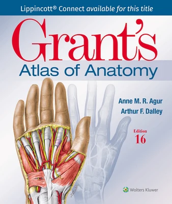
Illustrations drawn from real specimens, presented in surface-to-deep dissection sequence, set Grant's Atlas of Anatomy apart as the most accurate illustrated reference available for learning human anatomy and referencing in dissection lab. A recent edition featured re-colorization of the original Grant's Atlas images from high-resolution scans, also adding a new level of organ luminosity and tissue transparency. The dissection illustrations are supported by descriptive text legends with clinical insights, summary tables, orientation and schematic drawings, and medical imaging.
Author: Anne M. R. Agur,Arthur F. Dalley II
Publisher: LWW
Published: 02/06/2024
Pages: 896
Binding Type: Paperback
Weight: 5.09lbs
Size: 10.72h x 9.26w x 1.49d
ISBN: 9781975193430
This title is not returnable
- Renowned, high-resolution, dynamically colored illustrations organized in dissection sequence enable the formation of 3D constructs for each body region and provide detailed, realistic reference during dissection.
- Tables detail muscles, vessels, and other anatomic information in an easy-to-use format ideal for review and study.
- Enhanced medical imaging includes more than 100 clinically significant MRIs, CT images, ultrasound scans, and corresponding orientation drawings to help students confidently apply the laboratory experience to clinical rotations.
- Color schematic illustrations reinforce the relationships of structures and anatomical concepts in vibrant detail.
Author: Anne M. R. Agur,Arthur F. Dalley II
Publisher: LWW
Published: 02/06/2024
Pages: 896
Binding Type: Paperback
Weight: 5.09lbs
Size: 10.72h x 9.26w x 1.49d
ISBN: 9781975193430
This title is not returnable
Description
Illustrations drawn from real specimens, presented in surface-to-deep dissection sequence, set Grant's Atlas of Anatomy apart as the most accurate illustrated reference available for learning human anatomy and referencing in dissection lab. A recent edition featured re-colorization of the original Grant's Atlas images from high-resolution scans, also adding a new level of organ luminosity and tissue transparency. The dissection illustrations are supported by descriptive text legends with clinical insights, summary tables, orientation and schematic drawings, and medical imaging.
Author: Anne M. R. Agur,Arthur F. Dalley II
Publisher: LWW
Published: 02/06/2024
Pages: 896
Binding Type: Paperback
Weight: 5.09lbs
Size: 10.72h x 9.26w x 1.49d
ISBN: 9781975193430
This title is not returnable
- Renowned, high-resolution, dynamically colored illustrations organized in dissection sequence enable the formation of 3D constructs for each body region and provide detailed, realistic reference during dissection.
- Tables detail muscles, vessels, and other anatomic information in an easy-to-use format ideal for review and study.
- Enhanced medical imaging includes more than 100 clinically significant MRIs, CT images, ultrasound scans, and corresponding orientation drawings to help students confidently apply the laboratory experience to clinical rotations.
- Color schematic illustrations reinforce the relationships of structures and anatomical concepts in vibrant detail.
Author: Anne M. R. Agur,Arthur F. Dalley II
Publisher: LWW
Published: 02/06/2024
Pages: 896
Binding Type: Paperback
Weight: 5.09lbs
Size: 10.72h x 9.26w x 1.49d
ISBN: 9781975193430
This title is not returnable